Safe, Consistent Iron Delivery in Serum-Free Systems with Optiferrin® Recombinant Transferrin
Published on 30 June 2025
Application Note
EXECUTIVE SUMMARY
Efficient iron delivery is essential for cell growth and viability in vitro. Transferrin is widely used in cell culture media to fulfill this role. However, serum-derived transferrin poses challenges including batch variability, pathogen transmission risk, and regulatory complexity. InVitria’s Optiferrin is a recombinant human transferrin produced in a non-mammalian expression system that overcomes these limitations.
In this Application note, we show that Optiferrin performs equivalently to plasma-derived transferrin in iron delivery and hybridoma cell proliferation, validating its use in serum-free, animal-origin-free workflows.
Key Highlights
- Enables safe, receptor-mediated iron delivery
- Matches or exceeds performance of plasma-derived transferrin
- Supports proliferation in multiple cell types
- Facilitates serum-free, animal-origin-free, GMP-ready workflows
INTRODUCTION
Iron uptake is fundamental to cell metabolism, proliferation, and survival (Crescenzi et al., 2023). In living organisms, transferrin is the principal iron-binding protein in blood plasma. Human serum transferrin is a bilobal ~75 kD glycoprotein produced in the liver and found in blood plasma at approximately 2.5 mg/mL as a heterogeneous population: diferric (holo), monoferric N-lobe, monoferric C-lobe, or apo transferrin (Steere et al., 2012; Luck & Mason, 2012). Transferrin tightly binds Fe³+ ions and delivers them to cells via interaction with transferrin receptors (TFR) on the cell surface. Apo-transferrin has an extremely high affinity for ferric iron, with binding constants K′1 = 4.7 × 10²⁰ M-¹ and K′2 = 2.4 × 10¹⁹ M-¹. This ensures that apo-transferrin rapidly binds available iron in serum or culture media, quickly converting to holo-transferrin (Aisen et al., 1978). This receptor-mediated endocytosis ensures iron is supplied in a controlled, bioavailable form, supporting cellular processes while minimizing oxidative stress and toxicity from free iron (Luck & Mason, 2012; Lane et al., 2015).
In cell culture, transferrin is widely used as a supplement in serum-free and chemically defined media to mimic its physiological role in iron delivery (Luck & Mason, 2012). Historically, transferrin has been purified from human serum; however, this approach carries risks related to batch variability, pathogen transmission, and regulatory hurdles (Steere et al., 2012).
To address these limitations, recombinant human transferrin products such as Optiferrin have been developed using a non-mammalian expression system. Recombinant transferrin is structurally and functionally equivalent to plasma-derived transferrin, offering the same reversible iron binding, transferrin-receptor binding, and ability to promote cell proliferation and productivity.
RESULTS AND DISCUSSION
Optiferrin Delivers Bioavailable Iron via Receptor-Mediated Endocytosis
At physiological pH, transferrin binds Fe³+ and interacts with transferrin receptor 1 (TFR1) on the cell surface, initiating endocytosis and iron release within the endosome (Lane et al., 2015). This receptor-mediated process ensures controlled iron uptake and prevents oxidative stress from free iron in the medium (Kawabata, 2022).
Holo transferrin (diferric transferrin) exhibits the highest affinity for the cell surface transferrin receptor (TFR1), followed by the two monoferric forms (FeNhTF and FeChTF) (Luck & Mason, 2012). One of these three iron-bound forms of transferrin will bind the TFR1 and be internalized via clathrin-dependent endocytosis. The internalization of transferrin is rapid, with a half-life of approximately 3.5 minutes at 37°C (Steere et al., 2011). Subsequent acidification of the endosome through ATP-dependent H+ pumps triggers the receptor-stimulated release of iron from transferrin, where Fe³+ is reduced to Fe²+ before export to the cytosol (Luck & Mason, 2012).
The now iron-free transferrin/TFR1 complex is subsequently redirected back to the cell surface, where the weak association of apo transferrin for the TFR1 at serum pH leads to its dissociation, allowing apo transferrin to bind additional Fe³+ and repeat the cycle. Each full transferrin receptor cycle—from ligand binding to iron release and recycling—is completed in roughly 15–16 minutes, with about 90% of transferrin recycled within 30 minutes. A smaller portion follows slower recycling pathways, requiring up to 2 hours. These kinetics vary depending on cell type and conditions (Steere et al., 2011).
Although the fully saturated diferric transferrin has the highest affinity for the TFR (Kd ~4 nM), the fact that monoferric forms also are capable of high-affinity bonds with the TFR (Kd ~36 and 32 nM for the FeNhTF and FeChTF, respectively) would indicate that partially iron-saturated transferrin would be functional in cell culture (Luck & Mason, 2012).
These tightly regulated and rapid kinetics make transferrin a preferred and physiologically relevant source of iron in chemically defined media. Optiferrin has been shown to fully replicate this iron delivery mechanism. Its ability to bind TFR1 and participate in iron transport demonstrates functional parity with native, serum-derived transferrin.
![]()
Recombinant Optiferrin Retains Native Structure and Receptor Binding
Optiferrin is expressed at high levels in a non-mammalian expression system and has been rigorously analyzed to confirm the biochemical and functional aspects of this recombinant transferrin (Steere et al., 2012). Structural analysis verified that Optiferrin possesses the correct sequence and size and was able to compete with human serum-derived transferrin for TFR binding sites on both CCL-2 and Caco-2 cells (Zhang et al., 2012).
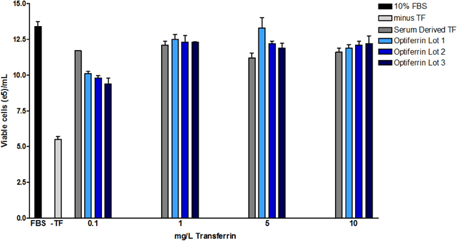
Optiferrin Supports Hybridoma Growth Comparable to Plasma-Derived Transferrin
Optiferrin demonstrated equivalent biological activity to native transferrin in supporting hybridoma cell proliferation across a relevant concentration range (0.1–10 mg/L). After 72 hours of culture in chemically defined conditions, viable cell counts were comparable between groups supplemented with Optiferrin and those with serum-derived transferrin.
Optiferrin Supports Stem Cell Differentiation and Maturation Through Efficient Iron Recycling and Delivery
Additionally, Dr. Paul Burridge’s laboratory at Northwestern University has employed Optiferrin in chemically defined media to support the differentiation and maturation of human induced pluripotent stem cells (hiPSCs), including cardiomyocytes. Their studies demonstrate that Optiferrin enables consistent iron delivery in xeno-free systems without compromising functional, metabolic, or transcriptional fidelity of derived cell types (Fetterman et al., 2024). Further, Burridge and colleagues have used Optiferrin in multiple iPSC-based models of cardiovascular disease and drug testing, underscoring its compatibility with high-performance, translational cell systems (Burridge et al., 2016; Sharma et al., 2017).
CONCLUSION
Optiferrin is a fully animal-origin-free, recombinant transferrin, making it ideally suited for workflows requiring stringent regulatory compliance and the elimination of adventitious agent risk. Its structural and functional equivalence to native human transferrin has been validated across a wide range of cell types—including hybridomas, mesenchymal stem cells, neural stem cells, induced pluripotent stem cells, T cells, fibroblasts, keratinocytes, VERO cells, and hematopoietic stem cells. This broad compatibility makes Optiferrin a highly versatile supplement for serum-free and chemically defined media.
As an animal-origin-free iron carrier, Optiferrin offers a consistent and scalable solution that supports robust cell proliferation, simplifies regulatory approval, and enables the transition to fully defined, xeno-free systems critical to modern biomanufacturing and cell therapy development.
MATERIALS AND METHODS
Sp2/0 hybridoma cells (ATCC) were used to assess the functional activity of recombinant transferrin in serum-free conditions. These cells are commonly used in antibody production workflows and serve as a relevant model for evaluating proliferation performance.
- Base medium: DMEM/F12
- Initial supplements:
- GlutaMax™ (ThermoFisher Scientific) – a stabilized dipeptide form of L-glutamine
- 10 mM HEPES – for buffering capacity
- 10% fetal bovine serum (FBS) – used during cell expansion before the assay setup
Cells were maintained under standard conditions (37°C, 5% CO₂) prior to assay.
Transferrin Bioactivity Assay Setup
To evaluate Optiferrin’s performance as a recombinant transferrin for serum-free media, hybridoma cells were transitioned to chemically defined conditions:
- Serum removal: Cells were washed thoroughly with basal DMEM/F12 to eliminate residual serum and prevent carryover of serum-derived transferrin.
- Defined media supplementation: Fresh basal medium was supplemented with:
- 1 g/L recombinant human albumin (rHSA) – for osmotic balance and carrier function
- 10 mg/L recombinant human insulin – to support glucose uptake and growth
- 6.7 µg/L sodium selenite – an essential trace element for enzymatic activity
- 2 mg/L ethanolamine – a phospholipid precursor for membrane integrity
This defined supplementation mimics standard serum-free formulations used in production environments.
Transferrin Treatment:
Cells were seeded into 96-well plates and treated with either:
- Serum-derived human transferrin (control group)
- Optiferrin – InVitria’s recombinant transferrin for iron delivery in serum-free media
- Dosage range: 0.1 to 10 mg/L
- Replicates: Triplicate wells per concentration
- Incubation period: 72 hours under standard growth conditions
- Endpoint: Viable cell concentration was assessed using trypan blue exclusion and manual or automated cell counting.
Featured Solution
Optiferrin – Recombinant Transferrin for Iron Delivery in Serum-Free Media
 Optiferrin is a recombinant, animal-origin-free human transferrin designed to replace plasma-derived transferrin in chemically defined media. It enables safe, efficient iron delivery via transferrin receptor-mediated endocytosis and supports robust proliferation across a wide range of mammalian cell types. Optiferrin eliminates the risk of adventitious agents and lot-to-lot variability associated with serum-derived transferrin, helping biomanufacturers transition to scalable, xeno-free workflows for cell therapy, gene therapy, and vaccine production.
Download the Full Application Note
The following content is gated. Please, subscribe to open access to it.
Footnotes
References
- Aisen, P., Leibman, A., & Zweier, J. (1978). Stoichiometric and site characteristics of the binding of iron to human transferrin. Journal of Biological Chemistry, 253(6), 1930–1937. https://doi.org/10.1016/S0021-9258(19)62337-9
- Crescenzi, E., Leonardi, A., & Pacifico, F. (2023). Iron metabolism in cancer and senescence: A cellular perspective. Biology, 12(7), 989. https://doi.org/10.3390/biology12070989
- Fetterman, K. A., Blancard, M., Lyra-Leite, D. M., Vanoye, C. G., Fonoudi, H., Jouni, M., DeKeyser, J. L., Lenny, B., Sapkota, Y., George, A. L., & Burridge, P. W. (2024). Independent compartmentalization of functional, metabolic, and transcriptional maturation of hiPSC-derived cardiomyocytes. Cell Reports, 43(5), 114160. https://doi.org/10.1016/j.celrep.2024.114160
- Kawabata, T. (2022). Iron-induced oxidative stress in human diseases. Cells, 11(14), 2152.https://doi.org/10.3390/cells11142152
- Lane, D. J. R., Merlot, A. M., Huang, M. L., Bae, D., Jansson, P. J., Sahni, S., Kalinowski, D. S., & Richardson, D. R. (2015). Cellular iron uptake, trafficking and metabolism: Key molecules and mechanisms and their roles in disease. Biochimica et Biophysica Acta (BBA) – Molecular Cell Research, 1853(5), 1130–1144. https://doi.org/10.1016/j.bbamcr.2015.01.021
- Luck, A. N., & Mason, A. B. (2012). Transferrin-mediated cellular iron delivery. In Current Topics in Membranes (Vol. 69, pp. 3–35). Academic Press. https://doi.org/10.1016/B978-0-12-394390-3.00001-X
- Sharma, A., Burridge, P. W., McKeithan, W. L., Serrano, R., Shukla, P., Sayed, N., Churko, J. M., Kitani, T., Wu, H., Holmgren, M., & Wu, J. C. (2017). High-throughput screening of tyrosine kinase inhibitor cardiotoxicity with human induced
pluripotent stem cells. Science Translational Medicine, 9(377), eaaf2584. https://doi.org/10.1126/scitranslmed.aaf2584 - Steere, A. N., Bobst, C. E., Zhang, D., Pettit, S. C., Kaltashov, I. A., Huang, N., & Mason, A. B. (2012). Biochemical and structural characterization of recombinant human serum transferrin from rice (Oryza sativa L.). Journal of Inorganic
Biochemistry, 116, 37–44. https://doi.org/10.1016/j.jinorgbio.2012.07.005 - Steere, A. N., Byrne, S. L., Chasteen, N. D., & Mason, A. B. (2011). Kinetics of iron release from transferrin bound to the transferrin receptor at endosomal pH. Biochimica et Biophysica Acta (BBA) – General Subjects, 1820(3), 326–333.
https://doi.org/10.1016/j.bbagen.2011.06.003 - Zhang, D., Lee, H., Pettit, S. C., Zaro, J. L., Huang, N., & Shen, W. (2012). Characterization of transferrin receptor-mediated endocytosis and cellular iron delivery of recombinant human serum transferrin from rice (Oryza sativa L.). BMC Biotechnology, 12, 92. https://doi.org/10.1186/1472-6750-12-92
- Burridge, P. W., Matsa, E., Shukla, P., Lin, Z. C., Churko, J. M., Ebert, A. D., Lan, F., Diecke, S., Huber, B., Mordwinkin, N. M., Neofytou, E., Paull, D., & Wu, J. C. (2016). Chemically defined generation of human cardiomyocytes. Nature Methods, 11(8), 855–860. https://doi.org/10.1038/nmeth.2999