


Improve Diagnostic Immunoassay Performance with Optibumin 25 Recombinant Human Serum Albumin
Published on 18 June 2026
Application Note
EXECUTIVE SUMMARY
Sensitive, reliable immunoassays are essential for early disease detection, where false-negative results can delay treatment. Albumin is commonly used in diagnostic immunoassays as a blocking agent and sample diluent, yet not all albumins perform the same. Aggregate populations present in conventional serum-derived albumins can scatter light and contribute to nonspecific background signal, elevating the limit of blank (LoB) and, in turn, the limit of detection (LoD). Because the intensity of scattered light scales with the sixth power of particle radius, even trace populations of larger aggregates can disproportionately affect assay sensitivity. We therefore reasoned that a monodisperse, aggregate-free albumin should directly translate into lower background and lower LoD.
Dynamic light scattering (DLS) confirmed that Optibumin 25 was monodisperse across the full 1-200 mg/mL concentration range evaluated, with PDI values below 0.1, a 4-7 nm hydrodynamic diameter, and no detectable light-scattering aggregates. In contrast, conventional diagnostic albumins, including another supplier’s recombinant HSA, were polydisperse and contained light-scattering aggregate populations. Consistent with this physical profile, when used as both an ELISA blocker and sample diluent for endogenous IL-6 detection in a human serum background, Optibumin 25 improved sensitivity 2.26-fold, reduced LoD by approximately 70% compared with diagnostic-grade BSA (0.53 vs 1.75 pg/mL), and improved dilution linearity.
Key Highlights
- PDI < 0.1 across 1-200 mg/mL by DLS
- No detectable light-scattering aggregate population
- Approximately 70% lower IL-6 LoD than diagnostic-grade BSA
- 2.26-fold improvement in assay sensitivity
- Improved dilution linearity in a human serum IL-6 ELISA
- Provides a recombinant, animal-origin-free alternative to serum-derived albumin and BSA
INTRODUCTION
Diagnostic procedures are critical for effective healthcare. Sensitive and reliable assays are necessary to detect health problems early and enable effective treatment; false-negative results can have dire consequences for patients sent home without treatment. In extreme cases, such as a failure to detect serum cardiac troponin I, a marker of myocardial infarction, the consequences can be fatal. Despite this, several widely used diagnostic procedures, including immunoassays such as ELISA, rely on animal-derived components like serum albumin that can introduce variability and reduce sensitivity by increasing nonspecific background signal.
While the use of albumin is ubiquitous in diagnostic immunoassays in the blocking step and in sample dilution, not all albumins perform the same (Xiao, Y., & Isaacs, S, 2012). The dominant physical differentiator is polydispersity. Conventional albumin preparations contain a broad distribution of particle sizes, including sub-visible oligomers and macro-particle aggregates. These aggregates prevent even, confluent coating of the microplate during blocking, leaving exposed surface that enables nonspecific binding in later steps; (Ma et al., 2020; CANDOR Bioscience GmbH, n.d.) when present in a sample diluent, they can also seed aggregation of serum proteins. Critically, these aggregates are potent light scatterers. Under the Rayleigh approximation the intensity of scattered light is proportional to the sixth power of particle radius, so a small mass fraction of macro-aggregates contributes disproportionately to scattered signal (Falke & Betzel, 2019). In absorbance-based assays such as an HRP–TMB ELISA, scattered light fails to reach the detector and registers as apparent absorbance, producing a false signal that elevates the LoB and therefore the LoD (Armbruster & Pry, 2008).
Because polydispersity is frequently invisible to traditional mass-based purity metrics, size-exclusion chromatography can filter out or under-report large species, inflating apparent monomer purity where an albumin can appear highly pure yet still carry the aggregate burden that antagonizes sensitivity (United States Pharmacopeia, 2017). This motivated a dispersity-first view of reagent quality: an albumin engineered to be monodisperse and free of light-scattering aggregates should lower assay background and detection limits by construction.
Impurities in albumin reagents, such as endotoxin or antibodies, can independently interfere with ELISA results, and non-specific binding or cross-reaction involving such contaminants, including cross-reactivity between sample proteins, blocking albumin, and enzyme-labeled detection reagents, can elevate background signal and reduce assay accuracy (Jiang et al., 2021). The extent of all of these effects varies lot-to-lot for serum-derived albumin, necessitating incoming-lot screening that costs time and money (Luo et al., 2023). To address these issues, InVitria has developed Optibumin 25, a chemically defined, recombinant, animal-origin-free albumin. By virtue of its recombinant nature, animal-free expression system, and high qualityhigh-quality standards, Optibumin is free of these confounders. Here we first characterize the dispersity of Optibumin 25 by DLS, then demonstrate that its monodisperse profile translates into enhanced sensitivity, a lower LoD, and improved linearity when Optibumin 25 is used as an ELISA blocker and sample diluent for a human serum sample, compared with a standard diagnostic-grade bovine serum albumin.
RESULTS AND DISCUSSION
Optibumin 25 Is Monodisperse and Free of Light-Scattering Aggregates
The polydispersity index (PDI) reported here is a dimensionless measure of the breadth of a particle-size distribution. It is derived from the DLS autocorrelation function as the square of the standard deviation of the distribution divided by the mean hydrodynamic radius, (SD/Rh)2. As a relative guide for proteins, a PDI below 0.1 indicates a monodisperse, narrowly distributed population; 0.1 to 0.2 reflects intermediate polydispersity with some aggregation; and values above approximately 0.3 indicate a highly polydisperse sample with significant aggregation.
To establish the physical basis for an expected sensitivity advantage, the dispersity of Optibumin 25 was characterized by DLS on the Uncle™(Unchained Labs) system across 1–200 mg/mL and compared with three conventional diagnostic albumins: a competitor’s diagnostic-grade recombinant HSA (rHSA), diagnostic-grade bovine serum albumin (BSA), and plasma-derived human serum albumin. At every concentration tested, Optibumin 25 exhibited a PDI below 0.1 (0.019–0.056), a mean hydrodynamic diameter of 4–7 nm, and no evidence of light-scattering aggregates, and can be interpreted as completely monodisperse (Figure 1, Table 1). In contrast, all three other albumins were polydisperse and carried light-scattering aggregate populations: diagnostic BSA (PDI 0.10–0.22), the competitor rHSA (PDI 0.25–0.34, mean diameter > 20 nm), and human serum albumin (PDI 0.19–0.37, mean diameter > 20 nm).
Notably, the competitor rHSA, a recombinant albumin like Optibumin 25, was among the most polydisperse materials tested, demonstrating that low polydispersity is a property of InVitria’s manufacturing process rather than of recombinant expression alone. The DLS intensity distributions make the difference visible: every conventional albumin shows a monomer / single-species peak near 5–7 nm accompanied by a secondary population of large, light-scattering aggregate species, whereas Optibumin 25 resolves as a single species with no detectable aggregate fraction (Figure 1A). At 50 mg/mL, the concentration used as the 5% blocking and diluent solution in the assays below, Optibumin 25 had a PDI of 0.026, versus 0.197 for diagnostic BSA, 0.270 for the competitor rHSA, and 0.360 for human serum albumin (Figure 1B).
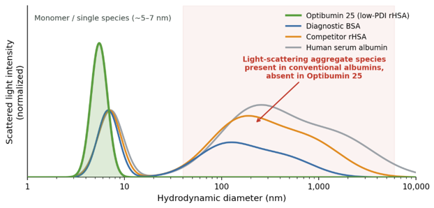

Table 1. DLS dispersity characterization of Optibumin 25 and comparator albumins.
DLS dispersity characterization of Optibumin 25 (low-PDI rHSA), a diagnostic-grade recombinant HSA (rHSA), diagnostic-grade bovine serum albumin (BSA), and plasma-derived human serum albumin across the indicated concentrations. PDI and mean hydrodynamic diameter were measured on the Uncle system.
| Albumin | Conc. (mg/mL) | PDI | PDI 95% CI |
| Optibumin 25 (low-PDI rHSA) | 200 | 0.06 | 0.008 |
| 100 | 0.03 | 0.004 | |
| 50 | 0.03 | 0.001 | |
| 10 | 0.02 | 0.012 | |
| 1 | 0.05 | 0.011 | |
| Competitor diagnostic rHSA | 200 | 0.34 | 0.046 |
| 100 | 0.25 | 0.085 | |
| 50 | 0.27 | 0.094 | |
| 10 | 0.32 | 0.013 | |
| 1 | 0.29 | 0.036 | |
| Diagnostic BSA | 200 | 0.15 | 0.092 |
| 100 | 0.22 | 0.033 | |
| 50 | 0.2 | 0.006 | |
| 10 | 0.11 | 0.019 | |
| 5 | 0.1 | 0.021 | |
| 1 | 0.1 | 0.025 | |
| Human Serum Albumin | 250 | 0.19 | 0.003 |
| 200 | 0.26 | 0.014 | |
| 100 | 0.34 | 0.018 | |
| 50 | 0.36 | 0.017 | |
| 10 | 0.37 | 0.017 | |
| 5 | 0.36 | 0.019 | |
| 1 | 0.37 | 0.019 |
Monodispersity Translates to Enhanced ELISA Sensitivity and a Lower Limit of Detection
To test whether the monodisperse profile of Optibumin 25 translates into improved assay performance, an HRP–TMB sandwich ELISA was performed using either Optibumin 25 or diagnostic-grade BSA as both the blocking agent and the sample diluent. Interleukin-6 (IL-6) was chosen as the model serum analyte because it is present at low levels in serum (pg/mL) and is a marker of numerous conditions, including respiratory infections, autoimmune disease, and cytokine storm, making it both challenging to detect and clinically relevant. A full procedure is provided in Materials & Methods. Briefly, plates were coated with capture antibody and blocked with 5% Optibumin 25 or 5% diagnostic BSA; serial dilutions of human AB serum were then prepared in the matching 5% albumin solution, and bound IL-6 was detected with a biotinylated matched detector antibody and streptavidin–HRP. The serum IL-6 concentration was determined to be 10.96 pg/mL by reference to a four-parameter logistic fit of a recombinant IL-6 standard curve run on the same plate.
The absorbance at 450 nm (A450) for each serum dilution was compared to a no-analyte control by one-way ANOVA with Dunnett’s post hoc test. Using a granular 2/3-fold dilution series, significant signal was detected in serum dilutions as low as 0.08× (0.96 pg/mL) in the Optibumin 25 group (Figure 2A) but only down to 0.19×(2.17 pg/mL) in the diagnostic BSA group (Figure 2B) representing a reproducible 2.26-fold improvement in sensitivity. The corresponding LoB, LoD, and LoQ are shown in Table 2. The LoD for the Optibumin 25 group was 0.53 pg/mL, whereas the LoD for diagnostic BSA was 1.75 pg/mL, a substantial ~70% reduction in the minimum detectable amount of IL-6. For analytes where a false negative carries clinical risk, a reduction of this magnitude can be consequential.
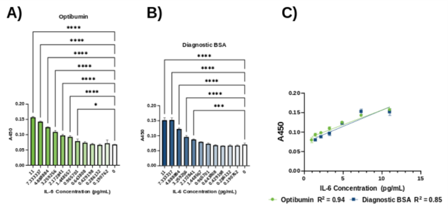
Table 2. LoB, LoD, and LoQ for IL-6 ELISA.
Limit of blank (LoB), limit of detection (LoD), and limit of quantification (LoQ) for the Optibumin 25 and diagnostic BSA groups corresponding to the results in Figure 2, expressed as IL-6 concentration (pg/mL).
| Parameter (pg/mL IL-6) | Optibumin 25 | Diagnostic BSA |
| Limit of Blank (LoB) | 0.27 | 1.62 |
| Limit of Detection (LoD) | 0.53 | 1.75 |
| Limit of Quantification (LoQ) | 2.01 | 2.04 |
Optibumin 25 Improves Dilution Linearity
With sufficient data points in the linear range, a linear regression was performed on the dilutions that were significantly different from their respective control (Figure 2C). The lines were similar, but the coefficient of determination, R2, was higher for Optibumin 25 than for diagnostic BSA (0.94 vs 0.85), indicating a more linear dilution response and thus more predictable quantification. Together with the lower LoD, this is the expected functional consequence of removing the polydisperse, light-scattering aggregate fraction: a cleaner baseline and a more uniform signal across the dilution series.
CONCLUSION
Reagent quality in high-sensitivity immunoassays is better described by dispersity than by mass-based purity alone. Optibumin 25 is monodisperse (PDI < 0.1) and free of the light-scattering macro-aggregates that elevate nonspecific background which is a property of InVitria’s manufacturing process rather than of recombinant origin alone, as a competing recombinant HSA evaluated here was itself polydisperse. As predicted by this physical profile, when used as an ELISA blocker and sample diluent, Optibumin 25 enhanced sensitivity up to 2.26-fold, reduced the IL-6 limit of detection by approximately 70%, and improved dilution linearity compared with diagnostic-grade BSA. Optibumin 25 is additionally free of the endotoxin and antibody contaminants known to confound ELISAs, and its recombinant, animal-origin-free manufacture delivers lot-to-lot consistency without incoming-lot screening. Taken together, the dispersity, performance, and quality characteristics of Optibumin 25 make it well suited as the albumin reagent for sensitive immunoassay applications.
MATERIALS AND METHODS
MaxSorp™ 96-well plates (ThermoFisher) were coated with 100 µL per well of a 1:1000 (v/v) dilution in PBS of IL-6 capture antibody (Abcam) for one hour at room temperature. Plates were decanted and blocked with 200 µL per well of either 5% (w/v) Optibumin 25 or 5% (w/v) diagnostic BSA overnight at 4 °C. The next day, serial dilutions of human AB serum were prepared in either 5% Optibumin 25 or 5% diagnostic BSA and transferred to a deep-well plate. The ELISA plate was decanted and 100 µL per well of sample was added for one hour at room temperature, with the albumin matched to the blocking condition. After incubation, the plate was decanted and washed three times with PBS + 0.05% (v/v) Tween® 20 (PBST). Plates were then treated with 100 µL per well of a 1:1000 (v/v) dilution of a biotinylated matched IL-6 detector antibody (Abcam), diluted in 1% (w/v) Optibumin 25 or diagnostic BSA in PBST. Plates were decanted, washed three times with PBST, and treated for 30 minutes with a 1:2400 (v/v) dilution of streptavidin–HRP conjugate in either 1% Optibumin 25 or 1% diagnostic BSA in PBST. Plates were decanted, washed three times with PBST, treated with TMB substrate, and developed in the dark for 30 minutes. The reaction was stopped with 0.16 M H2SO4 and absorbance at 450 nm was read on a microplate reader. DLS dispersity characterization (Figure 1, Table 1) was performed in triplicate on the Uncle system for 1–200 mg/mL protein solutions at 25 °C.
Featured Solution
Optibumin 25 – Recombinant Human Serum Albumin, 25% Solution – Animal-Origin-Free, GMP-Produced
Optibumin 25 is a recombinant human serum albumin developed for defined biomanufacturing and diagnostic workflows. In this application note, Optibumin 25 was evaluated as both an ELISA blocking agent and sample diluent for endogenous IL-6 detection in human serum.
Dynamic light scattering showed that Optibumin 25 was monodisperse across the tested concentration range, with no detectable light-scattering aggregate population. When used in the IL-6 ELISA workflow, Optibumin 25 reduced nonspecific background, lowered the limit of detection, and improved dilution linearity compared with diagnostic-grade BSA.
Ready to try Optibumin 25?
Get a free evaluation sample or request a quote for your project.
Frequently Asked Questions (FAQs)
What problem does this application note address?
This application note addresses nonspecific background and reduced sensitivity in diagnostic immunoassays. Albumin is commonly used as a blocking agent and sample diluent, but conventional albumins can contain aggregate populations that increase background signal and reduce assay performance.
Why does albumin quality matter in ELISA workflows?
Albumin can affect surface blocking, nonspecific binding, sample dilution, and background signal. If an albumin preparation contains aggregates or other interfering species, it may increase assay noise and make low-abundance analytes harder to detect.
What is polydispersity, and why is it important?
Polydispersity describes how broad the particle-size distribution is in a protein solution. A monodisperse albumin has a narrow, consistent size profile, while a polydisperse albumin contains a broader range of species, including aggregates. In sensitive assays, aggregates can contribute to light scattering and nonspecific background.
How was Optibumin 25 characterized in this study?
Optibumin® 25 was evaluated by dynamic light scattering across a 1–200 mg/mL concentration range. It showed PDI values below 0.1, a 4–7 nm hydrodynamic diameter, and no detectable light-scattering aggregate population.
How did Optibumin 25 compare with other albumins by DLS?
Optibumin 25 remained monodisperse across the tested concentration range. Diagnostic-grade BSA, plasma-derived human serum albumin, and a comparator recombinant HSA showed higher polydispersity and evidence of light-scattering aggregate populations.
What assay was used to test immunoassay performance?
The study used an HRP-TMB sandwich ELISA for endogenous IL-6 detection in human serum. Optibumin 25 or diagnostic-grade BSA was used as both the blocking agent and sample diluent.
What effect did Optibumin 25 have on IL-6 detection?
Optibumin 25 improved IL-6 assay sensitivity compared with diagnostic-grade BSA. The study showed a 2.26-fold improvement in sensitivity and an approximately 70% lower limit of detection.
What were the LoD results?
The IL-6 limit of detection was 0.53 pg/mL with Optibumin 25 and 1.75 pg/mL with diagnostic-grade BSA under the conditions tested.
Did Optibumin 25 improve dilution linearity?
Yes. Optibumin 25 improved dilution linearity in the human serum IL-6 ELISA, with an R² value of 0.94 compared with 0.85 for diagnostic-grade BSA.
Why might lower background improve diagnostic assay performance?
Lower background helps improve the separation between true analyte signal and assay noise. This can support lower detection limits and more reliable measurement of low-abundance biomarkers.
Is recombinant albumin automatically monodisperse?
No. The comparator recombinant HSA tested in this study was polydisperse. The data suggest that low polydispersity is not simply a result of recombinant origin, but is also tied to manufacturing process and product quality.
Why use Optibumin 25 instead of BSA or plasma-derived HSA?
Optibumin 25 is recombinant and animal-origin-free. It avoids reliance on bovine- or plasma-derived raw materials and provides a defined albumin option for immunoassay workflows where consistency, low background, and reliable detection are important.
Where can Optibumin 25 fit into diagnostic assay development?
Optibumin 25 may be evaluated as a blocking reagent, sample diluent component, or albumin-based assay stabilizer in ELISA and other immunoassay workflows where background reduction and reproducible low-level analyte detection are important.
The following content is gated. Please, subscribe to open access to it.
Footnotes
REFERENCES
- Armbruster, D. A., & Pry, T. (2008). Limit of blank, limit of detection and limit of quantitation. The Clinical Biochemist Reviews, 29 (Suppl 1), S49–S52. https://pmc.ncbi.nlm.nih.gov/articles/PMC2556583/
- CANDOR Bioscience GmbH. (n.d.). Surface blockers in serology: Some background about background [Technical note]. https://bocascientific.com/images/pdf/surface-blockers-in-serology-some-background-about-background.pdf
- Falke, S., & Betzel, C. (2019). Dynamic light scattering (DLS): Principles, perspectives, applications to biological samples. In A. S. Pereira, P. Tavares, & P. Limão-Vieira (Eds.), Radiation in bioanalysis: Spectroscopic techniques and theoretical methods (pp. 173–193). Springer. https://doi.org/10.1007/978-3-030-28247-9_6
- Ma, G. J., Ferhan, A. R., Jackman, J. A., & Cho, N. J. (2020). Conformational flexibility of fatty acid-free bovine serum albumin proteins enables superior antifouling coatings. Communications Materials, 1, Article 45. https://doi.org/10.1038/s43246-020-0047-9
- Jiang, X., Wu, M., Albo, J., & Rao, Q. (2021). Non-specific binding and cross-reaction of ELISA: A case study of porcine hemoglobin detection. Foods, 10(8), Article 1708. https://doi.org/10.3390/foods10081708
- Luo, Y., Pehrsson, M., Langholm, L., Karsdal, M., Bay-Jensen, A. C., & Sun, S. (2023). Lot-to-lot variance in immunoassays: Causes, consequences, and solutions. Diagnostics, 13(11), Article 1835. https://doi.org/10.3390/diagnostics13111835
- United States Pharmacopeia. (2017). Recombinant albumin human. In USP–NF. United States Pharmacopeial Convention.
- Xiao, Y., & Isaacs, S. N. (2012). Enzyme-linked immunosorbent assay (ELISA) and blocking with bovine serum albumin (BSA): Not all BSAs are alike. Journal of Immunological Methods, 384(1–2), 148–151. https://doi.org/10.1016/j.jim.2012.06.009