


Recombinant Albumin as a Carrier Protein Improves Cytokine Recovery and Functional Stability at Dilute Concentrations
Published on 12 January 2026
Application Note
Audio Summary: Optibumin 25 Stabilizes High-Cost Cytokines
EXECUTIVE SUMMARY
High-value cytokines and growth factors are the economic and functional drivers of modern cell therapy and biomanufacturing, frequently accounting for over 80% of total media costs. However, these critical investments are constantly at risk due to inherent protein fragility, leading to rapid aggregation, surface adsorption, and oxidative degradation that compromise process bioactivity and consistency. While plasma-derived human serum albumin (HSA) has historically been used to mitigate these losses, it introduces new liabilities regarding viral safety and lot-to-lot variability. To resolve these challenges, Optibumin® 25, a recombinant human serum albumin (rHSA), provides a superior, animal-origin-free solution that maximizes reagent recovery and unlocks room-temperature stability, safeguarding your process against cold-chain failures while eliminating the risks of serum-derived components.
Key Highlights
- Higher cytokine recovery at dilute concentrations
- Preserves cytokine bioactivity during storage
- Supports room-temperature stability for up to 4 weeks
- Comparable performance to plasma-derived HSA
- Animal-origin-free, recombinant formulation Improves recovery of dilute cytokine formulations by reducing aggregation and surface adsorption losses
INTRODUCTION
Cytokines are a diverse family of small, potent, secreted proteins that act as key signaling molecules in the immune system, coordinating the development, activation, and regulation of immune cells. They include diverse groups such as interleukins, interferons, and chemokines, and together these proteins orchestrate inflammation, host defense, and tissue homeostasis (Oppenheim, 2001). Some cytokines such as interleukin 2 (IL-2) and granulocyte-macrophage colony stimulating factor (GM-CSF) are used directly as therapeutics to modulate immune responses and tissue homeostasis in diseases such as cancer, autoimmune disorders, and chronic infections (Noble and Goa, 2012, Costello, 2009). Cytokines are also widely used in cell and gene therapy to control immune cell survival, expansion, and functional differentiation.
Growth factors are a class of proteins similar to and sometimes overlapping with cytokines that induce cell signaling events that initiate the cell cycle and drive cell expansion (Goustin et al., 1986). Without these proteins, cell biology research, vaccine manufacturing, in vitro drug screening, and advanced cell and gene therapies would not be possible because cells could not be expanded ex vivo.
Unfortunately, despite their vital roles in cell culture and therapeutic applications, the use of cytokines and growth factors presents inherent challenges. These proteins are very expensive relative to other cell culture components. For example, in Essential 8 (E8), a common iPSC media, over 80% of the cost is due to growth factors alone spurring leaders in the field to explore developing low-cost alternatives (Kuo et al., 2020). Additionally, due to the structure and typical use for these proteins, consistently maintaining highly bioactive samples is difficult.
Many cytokines and growth factors have a tendency to aggregate due to their hydrophobic and poorly soluble structures which frequently include helical bundle folds (Ricci and Brems, 2004). This aggregation reduces bioactivity and impacts consistency which disrupts cell culture applications (Roberts, 2015). Thus, their formulation presents one of the most significant challenges in biopharmaceutical development. Additionally, several therapeutic cytokines such as Interleukin-2 (IL-2), Interferon-beta (IFN-beta), and Insulin-like Growth Factor I (IGF-I) are characterized by extreme potency. This necessitates formulation at dilute microgram or nanogram concentrations to mitigate the risk of overdosing associated with administering low volumes of concentrated proteins (Lipiainen et al., 2014). At these dilutions, the physicochemical stability of the protein is compromised by rapid adsorption to container surfaces (Sonderby et al., 2018), increased tendency to form hydrophobic aggregates in response to agitation (Treuheit et al., 2002), and oxidative degradation mediated by reactive oxygen species (ROS) and trace metal ions in larger molar excess (Davies, 2016). Therefore, cytokines and growth factors are often formulated with carriers or excipients to improve their stability and recovery to maintain consistent bioactivity of these costly proteins.
Historically, human serum albumin (HSA) has been a commonly used excipient in cytokine therapeutics as well as a carrier in cytokine products used for cell and gene therapy workflows (Kato, 1987; Kwan, 1985; Yasushi et al. 1985; Lipiainen et al., 2014, Tarelli et al., 1998, Hawe and Friess, 2007). HSA reduces non-specific adsorption of cytokines to containers by creating a sacrificial monolayer on glass and plastic surfaces; moreover, HSA reduces denaturation caused by interaction with air-liquid interfaces (Ruiz et al., 2003, Tzannis et al., 1996, Visor et al., 1990, Velankar et al., 2024;). Through its free thiol residue (Cys34) and metal-binding N-terminus, HSA acts as a scavenger of peroxides and a chelator of Fenton-active metals (Taverna et al., 2013). Albumin exerts chaperone-like effects, binding transiently exposed hydrophobic regions to prevent aggregation and amyloid fibrillization, a critical function for hydrophobic cytokines (Finn et al., 2012). Altogether, these stabilizing attributes of HSA point toward its use as vital to increasing cytokine recovery, especially in low-concentration formulations. However, a historical barrier to albumin adoption is viral safety. Plasma-derived HSA requires rigorous donor screening and pasteurization (60°C for 10 hours), a process step. While effective, a residual risk of non-enveloped viruses or prions remains a regulatory consideration.
Other common excipients for cytokine and growth factor formulations include surfactants such as polysorbates and synthetic polymers such as polyethylene glycol (PEG). Either of these alternatives can reduce protein aggregation, but they have significant drawbacks compared to albumin. For example, polysorbate can cause peroxide formation (Kozuch et al., 2023) or particle formation due to oxidation and hydrolysis (Kim et al. 2022) while PEG often requires chemical modification of the protein of interest and has been associated with immunogenicity (Chen et al., 2021).
To meet the needs of cytokine and growth factor formulation and address the issues related to plasma-derived HSA, InVitria developed Optibumin 25, an animal-free, recombinant human serum albumin (rHSA). Optibumin 25 is manufactured to high quality standards to provide exceptional purity, monomer content, free-thiol content, stability, and consistency. Here we employ Optibumin 25 to improve stability of a cytokine widely used both as a therapeutic drug and a reagent in cell and gene therapy.
RESULTS AND DISCUSSION
Optibumin 25 as a Carrier Reduces Cytokine Aggregation and Improves Recovery
The impact of using Optibumin 25 and plasma-derived HSA as a carrier to reduce aggregation and improve thermal stability was evaluated. A commercially available cytokine was formulated in a relevant buffering system supplemented either with Optibumin 25, plasma-derived HSA or no albumin. Because cytokines are often formulated or suggested to be used and stored at or below 500 µg/mL levels, the cytokine was formulated at both 10 and 100 µg/mL concentrations for evaluation. The different cytokine formulations were incubated at 2–8°C, room temperature and 35–40°C for one month.
Analysis of cytokine concentration in the formulations at different time points was performed using a chemiluminescence based capillary system. The percentage of monomer recovery at different time points was calculated by measuring the concentration of monomer at a given time point divided by the concentration measured at Time-0.
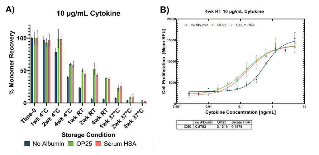
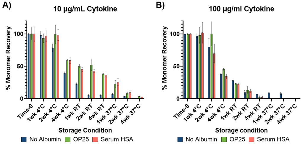
Results for formulated cytokine at a concentration of 10 µg/mL are shown in Figure 1A. The cytokine monomer recovery at 2–8°C remained stable for 1 week. The formulation with no HSA, recovery at 2–8°C began to decline steadily starting at 2 weeks; however, for the formulations containing Optibumin 25 and plasma-derived HSA, the stability extended to 2 weeks with recoveries at 99 and 98%, respectively. After 1 month at 2–8°C, all formulations decreased but the recovery of formulations with Optibumin 25 and HSA was about 20% higher compared to the formulation with no HSA. At room temperature, all formulations showed a marked decline to 50% or lower recovery after 1 week. After 2 weeks at room temperature, the formulation with no HSA fell to <10%, while the formulations with HSA maintained recoveries in the 40–50% range out to 4 weeks. Formulations incubated at 37°C showed similar trends as room temperature with overall low recovery but improved when Optibumin 25 or plasma-derived HSA is used.
Results for formulated cytokine at a concentration of 100 µg/mL are shown in Figure 1B. Similar to the lower cytokine concentration results, recovery of the cytokine at 2–8°C remained stable after 1 week; however, at this elevated cytokine concentration, at two weeks at 2–8°C only the formulation with Optibumin 25 provided stable recovery (~100%) compared to plasma-derived HSA (~70%) and no HSA (~80%). At room temperature and 37ºC, the albumin formulations showed no improvement on recovery.
It is worth noting that there was noticeable difference in albumin’s ability to stabilize and improve recovery of the cytokine at the two different cytokine concentrations. For formulations with 10 µg/mL cytokine concentration, albumin stabilized the cytokine in every condition; but at 100 µg/mL cytokine, albumin’s effect was muted. This observation could be attributed to two things albumin protects against: surface adsorption and shielding of hydrophobic regions. Given the same liquid to surface exposure (see storage details in Material and Methods), this loss and albumin’s prevention of it, would be more noticeable in the 10 µg/mL cytokine formulation.
The results here confirm Optibumin 25 as a potential carrier or excipient for cytokine formulation to improve stability and reduce loss to aggregation. These results also shed light on the complexity of protein formulation when working with hydrophobic proteins such as cytokines, by showing that even small adjustments to formulations can improve or reduce the impact of an excipient or carrier.
Notably, the stabilizing effect of albumin was concentration-dependent. At lower cytokine concentrations (10 µg/mL), inclusion of albumin substantially improved recovery across storage temperatures, consistent with mitigation of surface adsorption and aggregation losses. At higher cytokine concentrations (100 µg/mL), where surface-mediated losses represent a smaller fraction of total protein, the benefit of albumin inclusion was reduced under elevated temperature conditions.
Optibumin 25 as a Carrier Improves Functional Stability
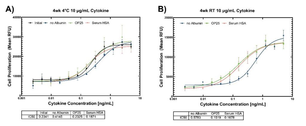
Reducing aggregation and improving stability are encouraging signs of an effective formulation of a cytokine or growth factor, but ultimately maintaining bioactivity is the principal goal of cytokine and growth factor formulation work. To assess bioactivity of the previously mentioned cytokine formulations, a bioassay was performed using a cell line that is well established to proliferate in response to the model cytokine/growth factor of interest. Briefly, cells were washed by centrifugation to remove serum growth factors then treated overnight with serial dilutions of several cytokine formulations evaluated in the previous aggregation experiment. These formulations included fresh cytokine at 10 µg/mL with no albumin, and cytokine at 10 µg/mL with either no albumin, plasma HSA or Optibumin 25 stored for 4 weeks at either 4°C or room temperature. The day after cell treatment, a thymidine analog was added to label only the proliferating cells. After incubation with the thymidine analog, cell samples were harvested and fixed, and the mean fluorescence intensity of the cell populations (a measure of cell proliferation) was characterized by flow cytometry.
The mean fluorescent intensity was then plotted against the log of the cytokine concentrations, and the data fitted to a four-parameter logistic curve (Figure 2). The EC50, the cytokine concentration needed to generate a half maximal growth response, was used to assess activity. A lower EC50 is indicative of higher cytokine activity because less cytokine is needed to generate a response in the cell population. In the 4°C conditions, (Figure 2A), the presence of albumin, either recombinant or plasma-derived, maintained cytokine activity comparable to fresh sample whereas in the group with no albumin the EC50 nearly doubled, meaning the activity was approximately halved. In the room temperature conditions, the same trend was observed but the formulation benefits of albumin, whether recombinant or plasma derived, was even more pronounced (Figure 2B). In these conditions, the EC50 of the albumin-free formulation increased to nearly four-fold that of the albumin containing conditions, meaning that the activity decreased four-fold. Interestingly, the albumin containing samples stored at room temperature maintained similar activity to those stored at 4°C and the fresh samples. This suggests that dilute cytokine solutions formulated with albumin maintain functional bioactivity for up to 4 weeks under room-temperature storage conditions evaluated in this study. This learning could improve the flexibility of cell therapy manufacturing processes that use these cytokines, eliminates concern about temperature excursion events, and could improve access to therapeutic cytokines in remote areas where cold storage is a challenge. Additionally, it is likely that similar benefits could be seen with additional proteins beyond cytokines.
Redox Properties of Optibumin 25 vs HSA
Oxidation is an insidious degradation pathway because an oxidized protein may remain soluble and monomeric but lose its biological potency or become immunogenic. While not explicitly evaluated in this study, the oxidation of cytokines is one of the key degradation pathways that is often addressed through formulation (Dion et al., 2018) or by protein design (e.g. mutation of a free cysteine) (An et al., 2024, Wang et al., 1984). Often small molecules such as L-methionine or ascorbate are used in formulations to slow oxidation, but these molecules have their
limitations. L-methionine is simply a sink for oxidants but cannot chelate ROS causing metals. Ascorbate can become pro-oxidant at certain concentrations in the presence of transition metals (Buettner and Jurkiewicz, 1996). Albumin is not limited in the same way as these small molecules. Due to its free thiol residue (Cys34) and high-affinity metal-binding N-terminus, albumin can scavenge free radicals and bind trace metals (Taverna et al., 2013), making it one of the most potent and useful antioxidants in cytokine formulation. In a previous application note titled Thiol by Combat: Enhanced Covalent Conjugation with Recombinant Albumin Compared to Human lasma-Derived Albumin, the free thiol content at Cys34 of Optibumin 25 was characterized and compared to serum-derived HSA. The key findings showed that across more than 10 lots of Optibumin 25 and 9 lots of serum-derived HSA, approximately 99% of Optibumin 25 remained in the reduced free-thiol form, compared to approximately 60% for serum-derived HSA. These results demonstrate that Optibumin 25 provides a higher proportion of redox-active albumin, a property relevant to mitigating oxidation-induced aggregation.
CONCLUSION
Cytokines and growth factors are high value proteins critical to clinical applications and cell-based research. However, they are unfortunately unstable at low concentrations due to aggregation, surface adsorption, and oxidation. Plasma albumin has historically been used to mitigate these stresses but has fallen out of favor due to concerns related to viral contamination and inconsistency. Here we have shown that Optibumin 25, a recombinant animal-free albumin, free from these concerns, can mitigate these stresses to reduce losses to aggregation and surface adsorption by approximately 20% at 2–8°C and up to 50% at room temperature under the dilute formulation and storage conditions evaluated which is comparable to plasma HSA. Furthermore, we have also demonstrated that Optibumin can maintain cytokine bioactivity for up to four weeks at room temperature. In practical cell culture workflows, improved functional stability of cytokines can translate to more consistent biological performance, reduced losses from aggregation or adsorption, and decreased need for compensatory over-dosing caused by activity decay over time. This enables short-term room-temperature storage of dilute cytokine formulations while maintaining functional activity, enhancing flexibility and robustness in cell therapy manufacturing and clinical cytokine storage. Given the mechanisms of albumin mediated stabilization, it is likely that these results would also be applicable to a wide range of other proteins such as antibodies and subunit vaccines.
MATERIALS & METHODS
Cytokine Formulation and Incubation
A bulk solution of cytokine at 200 µg/mL was prepared in a base buffer containing 2 mg/mL ascorbic free-acid and 23 mg/mL glycine, pH 4. From this bulk cytokine solution, the cytokine was diluted 1:1 in the base buffer containing either no albumin, 4 mg/mL Optibumin 25 or
4 mg/mL serum-derived HSA, such that the final cytokine concentration was 100 µg/mL and the final Optibumin 25 and serum HSA concentrations were 2 mg/mL. Cytokine formulations at 10 µg/mL were prepared by aliquoting from the 100 µg/mL formulations and diluting to 10-fold in the base buffer containing either no albumin, 2 mg/mL Optibumin 25 or 2 mg/mL serum-derived HSA. In one mL conical screw top tubes, one mL of the formulations were incubated for one month in a refrigerator maintained at 2–8°C, ambient room temperature, or an incubator at 35–40°C. Samples were taken for analysis at Time-0 and 1, 2, and 4 weeks.
Chemiluminescence Detection of Cytokine
Samples of each formulation were used for quantifying the concentration of cytokine monomer remaining after incubation at the various temperatures and time points. Cytokine monomer was detected using chemiluminescence based capillary electrophoresis on a ProteinSimple® Wes system (ProteinSimple, a Bio-Techne brand; San Jose, CA, USA). Samples were prepared under reduced and denatured conditions by reconstituting the fluorescent master mix supplied by ProteinSimple in DTT and SDS-containing sample buffer and mixing with the formulation samples at a 1 to 5 ratio (1 part fluorescent master mix and 5 parts diluted albumin samples). Cytokine monomer was analyzed using the Compass for SW software (version 6.3.0). The monomer area was quantified using dropped line peak analysis. For each formulation time point, the monomer area was divided by the initial measured concentration for that specific formulation at Time-0 using the Wes system and then multiplied by 100 to get a percentage. Each sample was run in duplicate, and the mean and standard deviation was calculated and reported.
Bioactivity Characterization by Cell Proliferation Assay
Formulations containing 10 µg/mL of cytokine stored at 4°C or room temperature were selected for the bioactivity assay due to the benefits of albumin inclusion on cytokine aggregation in these conditions. These formulations were diluted to a 10 ng/mL stock in cytokine-free cell culture media containing 5% fetal bovine serum (FBS). The stock was then serially diluted 2-fold twelve times. Next, cytokine responsive cells were harvested from culture media supplemented with 10% FBS and 10 ng/mL of cytokine. The cells were pelleted at 500 × g for 5 minutes, and the media were exchanged three times with cytokine-free assay media containing 5% FBS to remove any residual cytokine prior to the assay. The cells were aliquoted into the serially diluted cytokine at a 1:1 v/v ratio, making the highest concentration tested 5 ng/mL, and plated at 2 × 10⁴ cells per well in a 96-well plate for 16 hours. Then next day, 10 µM thymidine analog was added for 2 hours. The cells were then harvested by centrifugation, fixed in 4% paraformaldehyde for 15 minutes, and permeabilized in a saponin buffer for 20 minutes. Cell samples were stained with a thymidine analog detector conjugated to Alexa Fluor 647 and characterized by flow cytometry. Mean fluorescence intensity of each sample was plotted against the log of the concentration and fitted to a 4-parameter logistic fit to determine the EC50.
Featured Solution
Optibumin 25 – Recombinant Human Serum Albumin, 25% Solution – Animal-Origin-Free, GMP-Produced
Optibumin 25 is a chemically defined, recombinant human serum albumin designed to stabilize sensitive biologics, including cytokines and growth factors, used at low concentrations. Its high purity, high free-thiol (Cys34) content, and stabilizer-free formulation help reduce aggregation, adsorption, and oxidative degradation that compromise cytokine performance. As a plasma-free alternative to human serum albumin, Optibumin 25 supports more consistent cytokine recovery and preserved biological activity, while reducing regulatory and supply-chain risks associated with blood-derived materials. It is well suited for cell therapy, immunotherapy, and cytokine-dependent cell culture applications.
Ready to try Optibumin 25?
Get a free evaluation sample or request a quote for your project.
Frequently Asked Questions (FAQs)
Q: Why do cytokines lose activity at low concentrations?
A: Cytokines are highly potent proteins typically used at microgram- or nanogram-per-milliliter concentrations. At these dilutions, they are especially prone to surface adsorption, aggregation, and oxidative degradation, leading to reduced recovery and loss of biological activity over time.
Q: How does Optibumin® 25 improve cytokine stability?
A: Optibumin 25 acts as a carrier protein that reduces non-specific adsorption to containers, limits aggregation, and provides antioxidant protection through its free-thiol (Cys34) residue. This helps preserve both physical recovery and functional bioactivity of dilute cytokine formulations.
Q: How does recombinant albumin compare to plasma-derived HSA for cytokine stabilization?
A: In this study, Optibumin 25 performed comparably to plasma-derived HSA in preserving cytokine recovery and bioactivity, while eliminating donor-to-donor variability and residual viral risk associated with blood-derived materials.
Q: Does Optibumin® 25 support room-temperature storage of cytokines?
A: Yes. Cytokines formulated with Optibumin 25 maintained functional bioactivity for up to four weeks at room temperature under the conditions evaluated in this study, reducing sensitivity to handling and temperature excursions.
Q: What applications benefit most from cytokine stabilization with Optibumin® 25?
A: This approach is particularly relevant for cytokine-dependent workflows such as cell therapy manufacturing, T-cell immunotherapy, immune cell expansion, and other cell culture applications where consistency and bioactivity of cytokines are critical.
Download the Full Application Note

The following content is gated. Please, subscribe to open access to it.
Footnotes
References
- Oppenheim, J. J. (2001). Cytokines: Past, present, and future. International Journal of Hematology, 74(1), 3–8. https://doi.org/10.1007/BF02982543
- Noble, S., & Goa, K. L. (1997). Aldesleukin (recombinant interleukin-2). BioDrugs, 7(5), 394–422. https://doi.org/10.2165/00063030-199707050-00007
- Costello, R. T. (1993). Therapeutic use of granulocyte-macrophage colony-stimulating factor (GM-CSF): A review of recent experience. Acta Oncologica, 32(4), 403–408. https://doi.org/10.3109/02841869309093617
- Goustin, A. S., Leof, E. B., Shipley, G. D., & Moses, H. L. (1986). Growth factors and cancer. Cancer Research, 46(3), 1015–1029.
- Kuo, H.-H., Gao, X., DeKeyser, J.-M., Fetterman, K. A., Pinheiro, E. A., Weddle, C. J., Fonoudi, H., Orman, M. V., Romero-Tejeda, M., Jouni, M., Blancard, M., Magdy, T., Epting, C. L., George, A. L., Jr., & Burridge, P. W. (2020). Negligible-cost and weekend-free chemically defined human iPSC culture. Stem Cell Reports, 14(2), 256–270. https://doi.org/10.1016/j.stemcr.2019.12.007
- Ricci, M. S., & Brems, D. N. (2004). Common structural stability properties of 4-helical bundle cytokines: Possible physiological and pharmaceutical consequences. Current Pharmaceutical Design, 10(31), 3901–3911. https://doi.org/10.2174/1381612043382611
- Roberts, C. J. (2014). Therapeutic protein aggregation: Mechanisms, design, and control. Trends in Biotechnology, 32(7), 372–380. https://doi.org/10.1016/j.tibtech.2014.05.005
- Lipiäinen, T., Peltoniemi, M., Sarkhel, S., Yrjönen, T., Vuorela, H., Urtti, A., & Juppo, A. (2015). Formulation and stability of cytokine therapeutics. Journal of Pharmaceutical Sciences, 104(2), 307–326. https://doi.org/10.1002/jps.24243
- Sønderby, P., Bukrinski, J. T., Hebditch, M., Peters, G. H. J., Curtis, R. A., & Harris, P. (2018). Self-interaction of human serum albumin: A formulation perspective. ACS Omega, 3(11), 16105–16117. https://doi.org/10.1021/acsomega.8b02245
- Treuheit, M. J., Kosky, A. A., & Brems, D. N. (2002). Inverse relationship of protein concentration and aggregation. Pharmaceutical Research, 19(4), 511–516. https://doi.org/10.1023/A:1015108115452
- Davies, M. J. (2016). Protein oxidation and peroxidation. Biochemical Journal, 473(7), 805–825. https://doi.org/10.1042/BJ20151227
- Kato, Y., Hayakawa, E., Furuya, K., & Kondo, A. (1987). Method for solubilization of interferon (U.S. Patent No. 4,675,183). United States Patent and Trademark Office.
- Kwan, H. K. (1985). Biologically stable alpha-interferon formulations (U.S. Patent No. 4,496,537). United States Patent and Trademark Office.
- Yasushi, M., Kensuke, A., & Hajime, T. (1985). Stable composition of interleukin-2 and albumin (U.S. Patent No. 4,645,830). United States Patent and Trademark Office.
- Tarelli, E., Mire-Sluis, A., Tivnann, H. A., Bolgiano, B., Crane, D. T., Gee, C., Lemercinier, X., Athayde, M. L., Sutcliffe, N., Corran, P. H., & Rafferty, B. (1998). Recombinant human albumin as a stabilizer for biological materials and for the preparation of international reference reagents. Biologicals, 26(4), 331–346. https://doi.org/10.1006/biol.1998.0163
- Hawe, A., & Friess, W. (2007). Stabilization of a hydrophobic recombinant cytokine by human serum albumin. Journal of Pharmaceutical Sciences, 96(11), 2987–2999. https://doi.org/10.1002/jps.20909
- Ruiz, L., Reyes, N., Duany, L., Franco, A., Aroche, K., & Hardy Rando, E. (2003). Long-term stabilization of recombinant human interferon alpha 2b in aqueous solution without serum albumin. International Journal of Pharmaceutics, 264(1), 57–72. https://doi.org/10.1016/S0378-5173(03)00388-0
- Tzannis, S. T., Hrushesky, W. J. M., Wood, P. A., & Przybycien, T. M. (1996). Irreversible inactivation of interleukin-2 in a pump-based delivery environment. Proceedings of the National Academy of Sciences of the United States of America, 93(11), 5460–5465. https://doi.org/10.1073/pnas.93.11.5460
- Visor, G. C., Tsai, K. P., Duffy, J., Calderwood, T., & Knepp, V. M. (1990). Quantitative evaluation of the stability and delivery of interleukin-1β by infusion. Journal of Pharmaceutical Science and Technology, 44(3), 130–137. https://pubmed.ncbi.nlm.nih.gov/2370622/
- Velankar, K. Y., Gawalt, E. S., Wen, Y., & Meng, W. S. (2024). Pharmaceutical proteins at the interfaces and the role of albumin. Biotechnology Progress, 40(5), e3474. https://doi.org/10.1002/btpr.3474
- Taverna, M., Marie, A.-L., Mira, J.-P., & Guidet, B. (2013). Specific antioxidant properties of human serum albumin. Annals of Intensive Care, 3(1), Article 4. https://doi.org/10.1186/2110-5820-3-4
- Finn, T. E., Nunez, A. C., Sunde, M., & Easterbrook-Smith, S. B. (2012). Serum albumin prevents protein aggregation and amyloid formation and retains chaperone-like activity in the presence of physiological ligands. Journal of Biological Chemistry, 287(25), 21530–21540. https://doi.org/10.1074/jbc.M112.372961
- Kozuch, B., Weber, J., Buske, J., Mäder, K., Garidel, P., & Diederichs, T. (2023). Comparative stability study of polysorbate 20 and polysorbate 80 related to oxidative degradation. Pharmaceutics, 15(9), 2332. https://doi.org/10.3390/pharmaceutics15092332
- Kim, D. M., Stansberry-Perkins, K., Marlow, M. S., & Pyles, E. A. (2022). Human serum albumin mitigates formation of fatty acid particles in polysorbate-containing solutions. Journal of Pharmaceutical Sciences, 111(11), 3185–3188. https://doi.org/10.1016/j.xphs.2022.08.012
- Chen, B.-M., Cheng, T.-L., & Roffler, S. R. (2021). Polyethylene glycol immunogenicity: Theoretical, clinical, and practical aspects of anti-polyethylene glycol antibodies. ACS Nano, 15(9), 14022–14048. https://doi.org/10.1021/acsnano.1c05922
- Dion, M. Z., Leiske, D., Sharma, V. K., Zuch de Zafra, C. L., & Salisbury, C. M. (2018). Mitigation of oxidation in therapeutic antibody formulations: A biochemical efficacy and safety evaluation of N-acetyl-tryptophan and L-methionine. Pharmaceutical Research, 35(11), Article 222. https://doi.org/10.1007/s11095-018-2467-5
- An, Y. J., Jung, Y.-E., Lee, K. W., Kaushal, P., Ko, I. Y., Shin, S. M., Ji, S., Yu, W., Lee, C., Lee, W.-K., Cha, K., Lee, J.-H., Cha, S.-S., & Yim, H.-S. (2024). Structural and biochemical investigation into stable FGF2 mutants with novel mutation sites and hydrophobic replacements for surface-exposed cysteines. PLOS ONE, 19(9), e0307499. https://doi.org/10.1371/journal.pone.0307499
- Wang, A., Lu, S.-D., & Mark, D. F. (1984). Site-specific mutagenesis of the human interleukin-2 gene: Structure–function analysis of the cysteine residues. Science, 224(4656), 1431–1433. https://doi.org/10.1126/science.6427925
- Buettner, G. R., & Jurkiewicz, B. A. (1996). Catalytic metals, ascorbate and free radicals: Combinations to avoid. Radiation Research, 145(5), 532–541. https://pubmed.ncbi.nlm.nih.gov/8619018/